Homogeneous Immunoassay Construction Kit
ONEPot Immunoassay Kit <OpenGUS Method>
Highlights
-
Antigens are sandwiched and detected using two antibodies of your choice.
-
Unlike ELISA, there is no need to immobilize antibodies on plates or wash wells.
-
Just by mixing, the antibodies and recombinant GUS will form an affinity bond.
-
Proteins that stably form multimers may be possible to be detected with only one type of antibody.
-
Fluorescence measurement kit (Cat. #DS850) and absorption measurement kit (Cat. #DS860) are avilable.
Product information
This is a kit for constructing an homogenous immunoassay using your antibodies, based on the OpenGUS Immunoassay by mutant β-glucuronidase (GUS).
This product is commercialized by BDL based on the research results of Dr. Hiroshi Ueda, Dr. Tetsuya Kitaguchi, Dr. Bo Zhu and co-workers of Tokyo Institute of Technology (at the time of research)
Notes
– We recommend samples containing 3 μg/ml or more of the target protein.
(Assuming that the sample is diluted 100 times and target protein is measured as 30 ng/ml)
– The pair of mouse IgG1 that can sandwich antigens are required.
(When the antigen forms a multimer, there are cases where the antigen could be detected using only one type of antibody.)
– Signal intensity can be significantly affected by the composition of the sample solution.
– Detection sensitivity varies depending on antigen and antibody.
– Samples containing a large amount of immunoglobulin such as human serum/plasma/blood cannot be used because the background will be high.
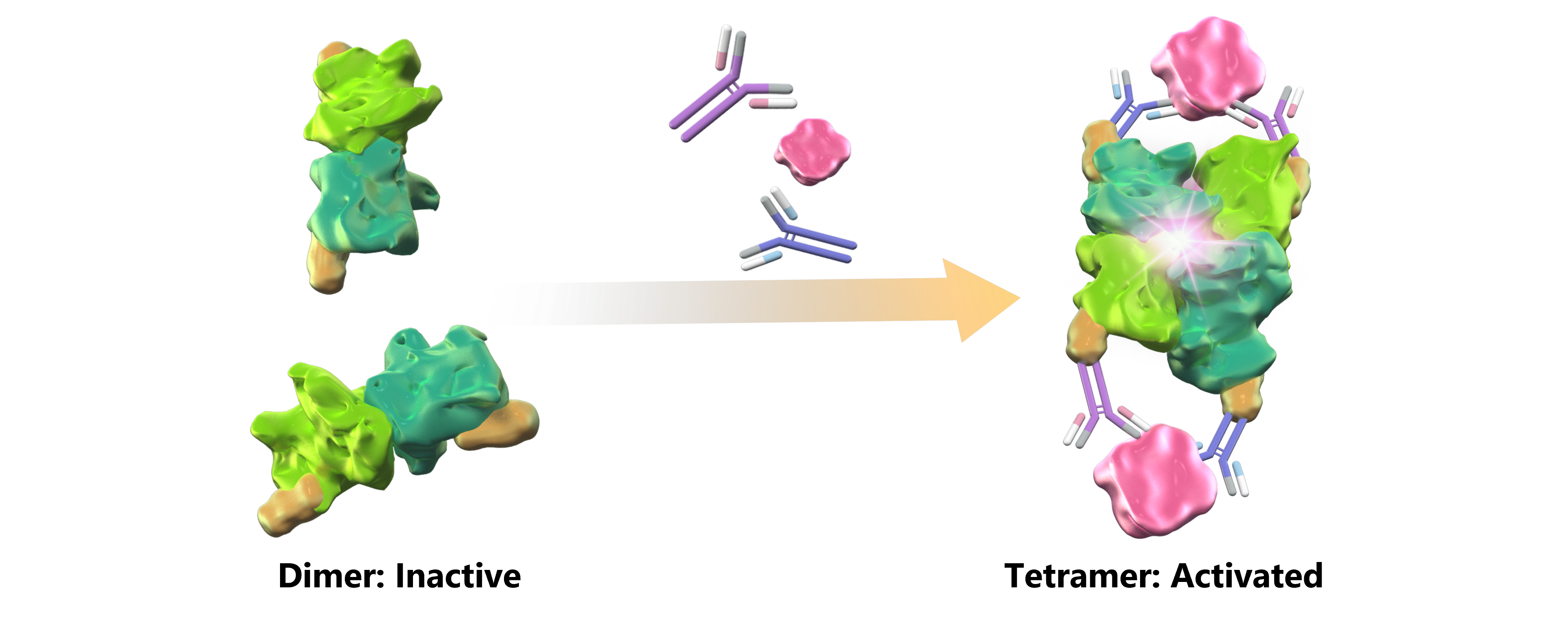
OpenGUS technology
β-glucuronidase (GUS) is an enzyme that hydrolyzes β-glucuronide to β-glucuronic acid. E. coli GUS exhibits activity by forming a tetramer. The mutant GUS used in this product has reduced affinity between monomers, and although it forms dimers, it rarely forms tetramers.
Recombinant GUS mixture, a component of this product, is a mutant GUS fused with antibody binding domains. Dimers of mutant GUS link together through antigen-antibody reactions and forcefully form tetramers. By measuring the activity of GUS, antigen concentration is estimated.
Procedure overview
1) Mix antigen, antibody and enzyme included in the kit
2) Incubate for 60 minutes
3) Add Substrate
4) Incubate for 60 minutes
5) Measure fluorescence or absorbance
Procedure overview
– Mouse IgG1 antibody pair that can sandwiching antigens
– Purified antigen for standard curve
– 96 well plate
– Microplate reader
Fluorescence (Cat. #DS850): Excitation 340 nm / Emission 450 nm
Absorbance (Cat. #DS860): Main wavelength 405nm / Reference 660nm
– Multichannel pipettor
Application 1 Measurement of Lactoferrin concentration in saliva and tears
Lactoferrin in saliva and tears was measured using this product. The results were compared with those obtained using the ELISA kit.
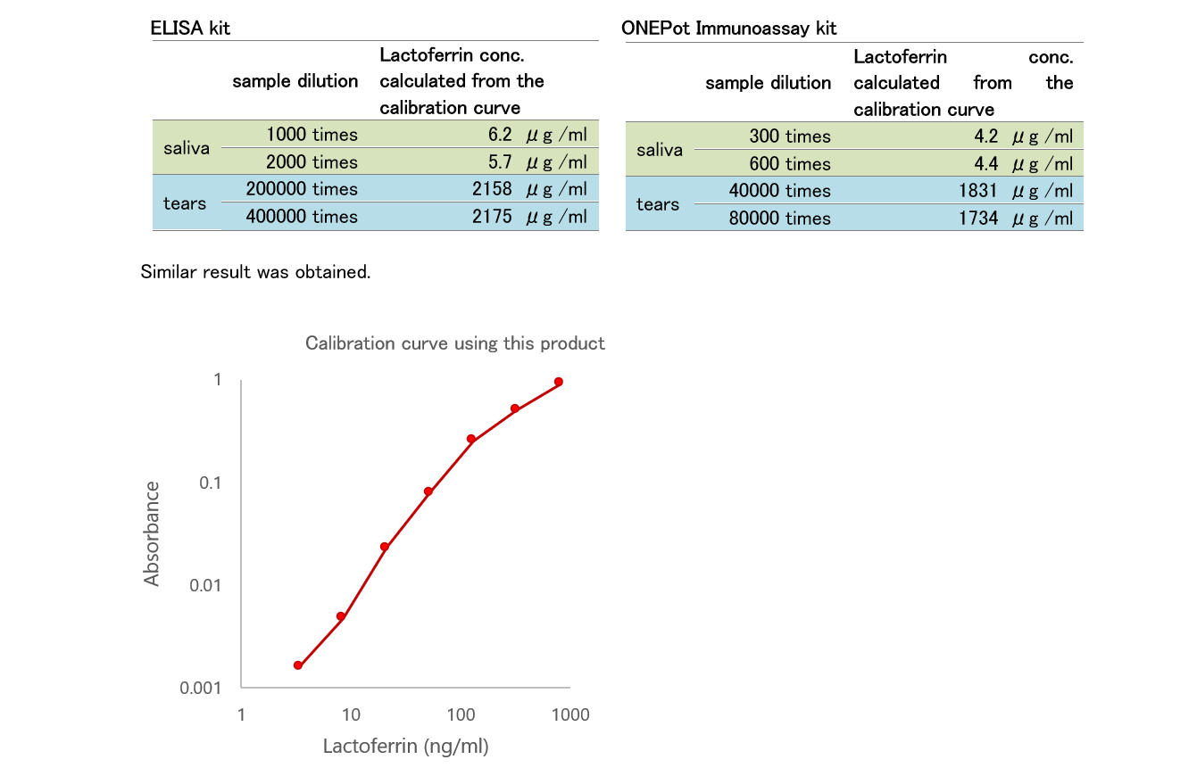
Antibody: Anti-Lactoferrin, Human, Mouse-Mono(1A1) (HyTest, #4L2-1A1)
*Detection was performed using only one type of antibody. Since Lactoferrin forms a multimer, we believe that only one type of antibody was required to detect Lactoferrin.
*The vertical axis of the calibration curve is the [Signal – Background] value
Application 2 Calibration curve obtained using purified Cry j 1 (cedar pollen antigen)
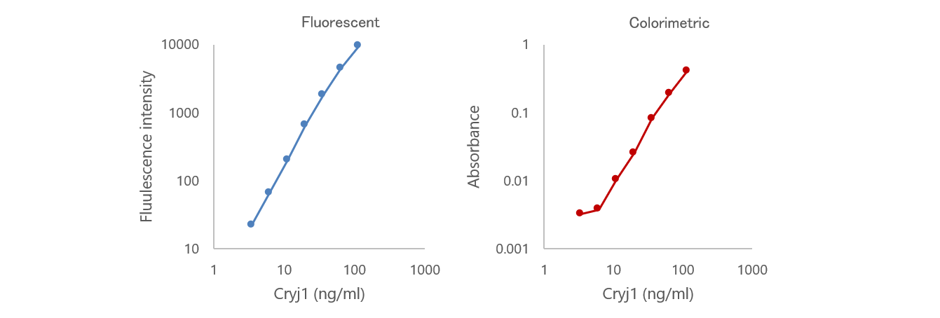
Antibodies: Anti-Cry j1 Mouse-Mono(013) (BDL #HBL-Ab-1-013) and Anti-Cry j1 Mouse-Mono(053) (BDL #HBL-Ab-1-053)
* Cry j 1 could be measured from Cry j 1 enriched samples, but the sensitivity was low to detect Cry j 1 from environmental samples.
*The vertical axis of the calibration curve is the [Signal – Background] value
Application 3 Calibration curve obtained using AAV2 (empty capsids)
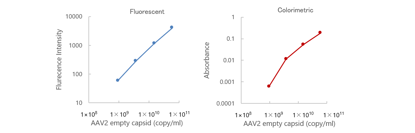
Antigen: AAV2 empty capsids (PROGEN #66V020)
Antibody: Anti-AAV2 (intact particle) mouse recombinant, (A20R) (PROGEN #610298)
* Detection was performed using only one type of antibody. Since antigens aligned on the Capsid surface, we believe that only one type of antibody was required to detect AAV empty capsids.
*The vertical axis of the calibration curve is the [Signal – Background] value
In addition, we have applications for human C-Reactive Protein (CRP) and Calf-Intestinal Alkaline Phosphatase (CIAP). Please see the product manual.
References
1) J. Su, D. Jinhua, T. Kitaguchi, Y. Ohmuro-Matsuyama and H. Ueda. Noncompetitive homogeneous immunodetection of small molecules based on beta-glucuronidase complementation. Analyst 143, 2096-2101 (2018)
2) J. Su, C. Beh, Y. Ohmuro-Matsuyama, T. Kitaguchi, S. Hoon and H. Ueda. Creation of stable and strictly regulated enzyme switch for signal-on immunodetection of various small antigens. J. Biosci. Bioeng. 128, 677-682 (2019)
3) J. Su, T. Kitaguchi, Y. Ohmuro-Matsuyama, T. Seah, F.J. Ghadessy, S. Hoon and H. Ueda. Transmembrane signaling on a protocell: Creation of receptor-enzyme chimeras for immunodetection of specific antibodies and antigens. Sci. Rep. 9, 18189 (2019)
4) B. Zhu, C. Qian, H. Tang, T. Kitaguchi and H. Ueda. Creating a thermostable beta-glucuronidase switch for homogeneous immunoassay by disruption of conserved salt bridges at diagonal interfaces. Biochemistry 62, 309–317 (2023)
5) B. Zhu, Y. Yamasaki, T. Yasuda, C. Qian, Z. Qiu, M. Nagamine, H. Ueda and T. Kitaguchi. Customizable OpenGUS immunoassay: a homogeneous detection system using β-glucuronidase switch and label-free antibody. Biosens. Bioelectron. 267, 116796 (2025)
This product is for research use only.
This product has been commercialized under the license from Institute of Science Tokyo.
This product or any modifications thereof may not be resold to any third party, used to manufacture commercial products, or provide services without the prior written consent of BioDynamics Laboratory Inc.
Code
Product name
size
1 Kit (for 100 well)
Manuals

SDS
–
Code
size
1 Kit (for 100 well)
Manuals
SDS
–
BDL is a member of the Funakoshi Group.